The erythrocytes or red blood cells are the most common cell type in the blood. In fact, they are the most common cell in the entire body. But let’s stick with the blood for now. Let’s look at its structure and function in the body.
Topic Outline:
There are 20 to 30 TRILLION red blood cells in an adult human. That’s trillion with a T. That’s a whole lot of cells!
And as we looked at in the last video of this series, they develop from a type of cell called reticulocytes. We’re not going to go through how they develop in this video, but if you want to see how awesome that process is, check out that video.
Red Blood Cell Structure
Now let’s take a look at the structure of these Red blood cells.
Red Blood Cells are Biconcave Disks
First off – Red blood cells have a very interesting shape. They have the shape of a biconcave disk. When something is concave, it curves inwards. And, that’s exactly what we see with red blood cells, except, we see it on both sides of the cell hence the term “biconcave”.

Now there are huge benefits to this. The first one is that it makes them small. This is great because while yes – there ARE some large blood vessels in the body, there are also MANY MORE very itsy bitsy tiny little blood vessels called capillaries that these cells have to go through to supply blood to places that need it.
The second reason is that it gives them a larger surface area to volume ratio. In other words, it’s gonna make it easier for things like Oxygen and carbon dioxide to get into and out of the cell.
Think about it like this – let’s say you have oxygen in the middle of a cell that has the shape of a sphere. It has a longer distance to travel to get to the cell membrane just so that it can cross into the surrounding tissues.
But, since you have this biconcave shape, it’s like the cell membrane is just right there. Whenever that oxygen needs to get out or in, it can more easily just go. It’s a beautiful thing.
Red Blood Cell Membranes have Spectrin
There’s also something else that’s very interesting about the structure of red blood cells – specifically the cell membrane. Their cell membrane has some specialized proteins – one of which is called spectrin.
What’s cool about this protein (and others like it) – is that it makes the cell membrane flexible. That comes in handy when it comes to getting into some tight spots. The cell is basically able to fold over itself just to fit into those tight squeezes.

Red Blood Cells Lack Cellular Organelles and Structures
And ANOTHER interesting feature is that if you compare a typical cell to a red blood cell, you’ll notice that there are a few things missing. Yep, it lacks key structures and organelles. For example, there’s no nucleus which also means that there’s no DNA inside a red blood cell.

It also doesn’t have any endoplasmic reticulum – this organelle is used to make proteins. So – we’re not making any proteins inside the red blood cells.
It also has no mitochondria. Mitochondria use oxygen to make ATP – which is the energy currency of the body. So – we’re not using up any of that oxygen that the red blood cells carry, which is ALSO a good thing.
Here’s the thing – Red Blood cells have one MAIN function – gas exchange. We want to deliver oxygen and we want to get rid of Carbon dioxide. And our red blood cells are specifically designed to accomplish this.
All that other stuff is unnecessary. So when they’re developing, they get rid of all that extra baggage. In fact, there’s a lot we can learn about life from these red blood cells. If you’re carrying around a bunch of unnecessary baggage, get rid of it.
But I digress.
The Hemoglobin Molecule in Red Blood Cells
Hemoglobin Structure
The hemoglobin molecule is probably the most important molecule in Red Blood Cells. This is a large molecule that’s made up of four protein molecules called globins. These four globins are alpha 1, alpha 2, beta 1, and beta 2.
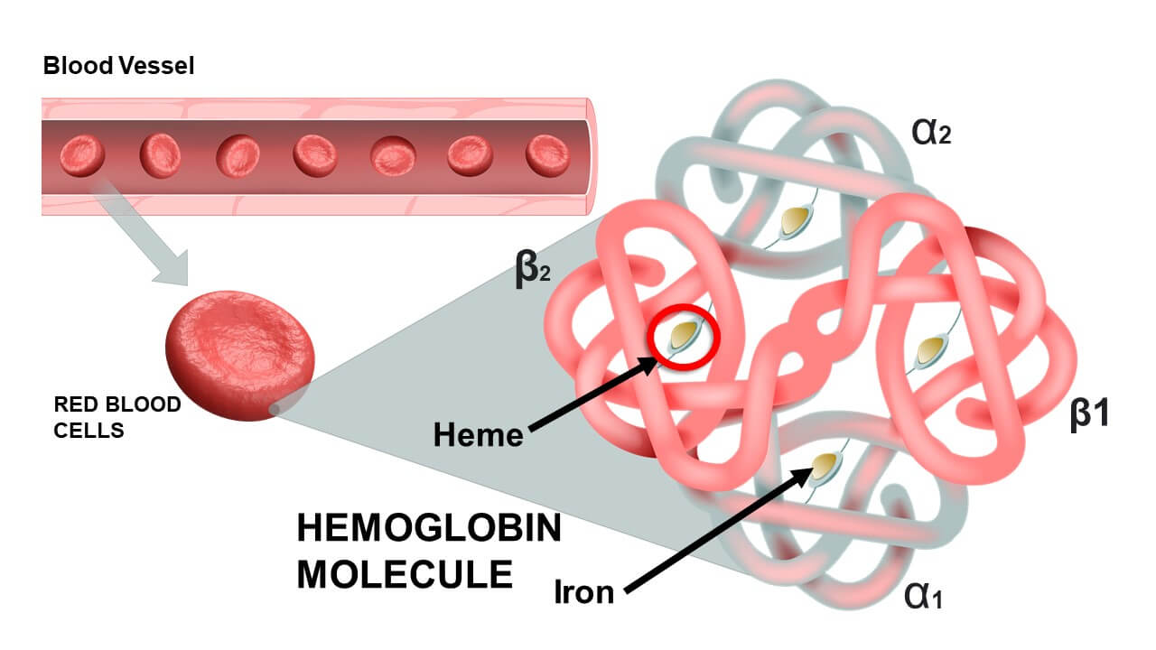
Each of these globins has a red pigment molecule bound to it and that molecule is called heme. And these heme molecules have one ion of iron.
Role of Hemoglobin in Oxygen Transport
Now, this is the important part – each iron molecule can hold onto one Oxygen molecule. And THAT is why we want these red blood cells in the first place.
Ok, let’s do some math here. Each heme molecule can hold onto 1 Oxygen molecule. Each hemoglobin has 4 heme molecules, which means that ONE hemoglobin molecule can carry up to 4 oxygen molecules.
Here’s where it gets crazy – One red blood cell – just one – contains about 300 million hemoglobin molecules, which means that ONE red blood cell can transport about 1.2 billion Oxygen molecules! That’s billion – like with a B. That’s a whole lot of oxygen.

What happens during gas exchange?
Formation of Oxyhemoglobin
When red blood cells are in the lungs, there’s going to be a lot of Oxygen available and the hemoglobin will latch onto the oxygen to become oxyhemoglobin. Oxyhemoglobin is bright red in color and that’s why the blood shows up bright red when it’s exposed to oxygen.
When red blood cells get to the other tissues, especially ones that are in need of oxygen, the hemoglobin’s affinity for oxygen will actually go down. There’s a structural change that happens in the hemoglobin molecule that makes it less likely to hold onto the oxygen.
This is good news because it causes the hemoglobin molecules to actually release the oxygen making them available for the tissues.
Now, this is actually a detailed process that I can’t go into fully right here, but I do have another video on this more as well as the idea of the Bohr effect. You can check that video here.
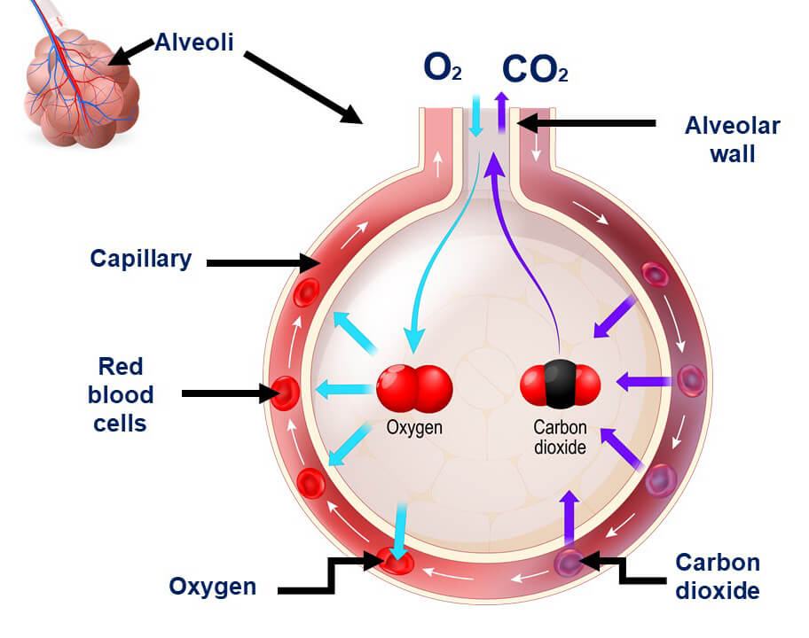
Formation of Carbaminohemoglobin
There’s one more thing I want to mention briefly and it has to do with Carbon dioxide. Cells use oxygen in the process known as cellular respiration. And one of the products of cellular respiration is carbon dioxide. That carbon dioxide needs to be dealt with because it can cause serious complications in the body.
Well, what we see is that:
- 7% of that Carbon dioxide gets carried in the blood as dissolved carbon dioxide
- 70% of it is converted to bicarbonate via an enzyme called carbonic anhydrase
- And finally, 23% of the carbon dioxide will bind to the hemoglobin to form carbaminohemoglobin.
Yes – hemoglobin plays a role even with carbon dioxide.
And of course, the blood travels back to the lungs where the hemoglobin in the red blood cells can release the carbon dioxide into the lungs so that you can breathe it out and the plants in your environment can use it up. It’s a beautiful thing.
Summary
We have learned that Red Blood Cells a.k.a. erythrocytes have the following structures:
- They are biconcave in shape.
- They have flexible membranes due to specialized proteins, specifically spectrin.
- They lack basic organelles and cellular structures.
We have also learned about the structure and importance of the hemoglobin structure in red blood cells.
In the next video, we’re going to talk about disorders that can happen with erythrocytes – what happens when things go wrong? Let’s read more…
Infographic
