This post was updated on March 24, 2022
Click here to leave a comment.
Ever wonder what happens in a heartbeat? What happens inside our heart when we hear the ‘lub-dub’ sound? When I say cardiac cycle, I’m talking about everything that happens from the beginning of one heartbeat to the beginning of the next. So we’re dealing with an entire heartbeat.
Introduction
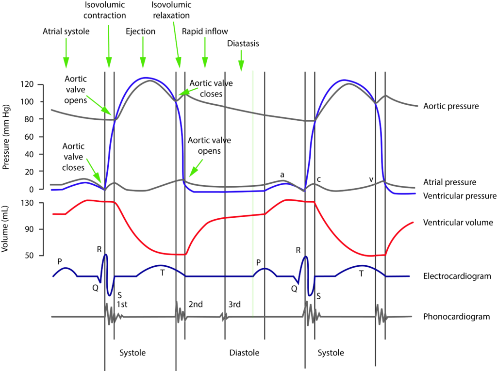
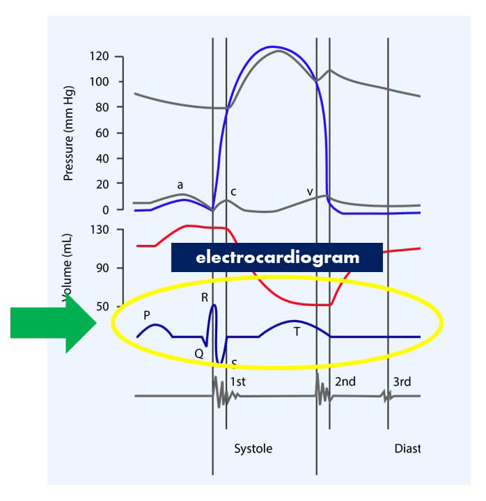
Let’s look at the cardiac cycle diagram. I know, it looks hard, but it isn’t really. Here’s why it seems so difficult. Because when your professor teaches you about the cardiac cycle, you see this complicated diagram right here.
It has a TON of details and it looks kinda scary. Let’s not do it like that. Let’s break it apart, and build it together.
What happens in a Cardiac Cycle?
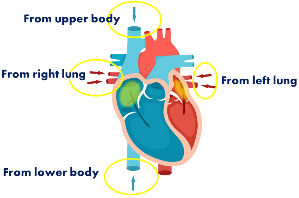
Let’s start with blood coming back from the body. Blood enters the heart first through the atria. On the left side, we have blood coming back from the lungs and on the right side, we have blood coming back from the rest of the body.
When the atria contract, they push blood into the ventricles. And then when the ventricles contract, they push blood out of the heart. Those are the two contractions that cause blood flow.
If this seems unfamiliar or unclear for you, head on to my article on How Blood Flows Through the Heart and you’ll get a more detailed explanation.
The Cardiac Cycle Diagram Explained
The first place we’re going to look at is the electrocardiogram. We’re going to start here because this shows the electrical signals that are responsible for the heart beating.
Atrial Systole
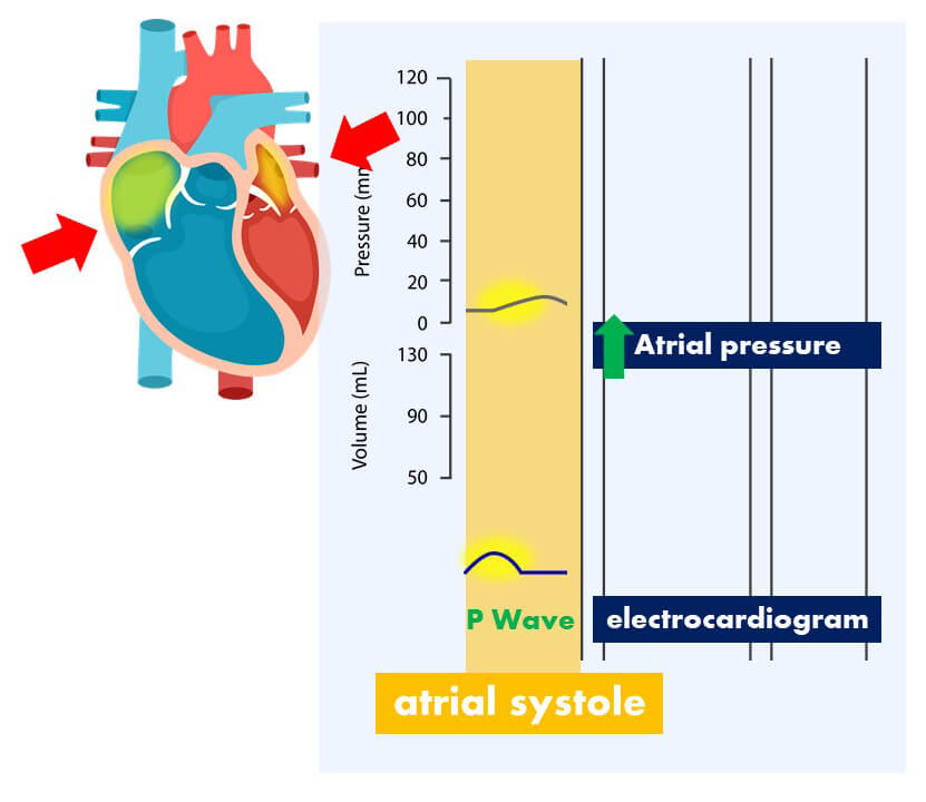
The first thing we see is the P wave. This shows the depolarization of the atria. That’s the electrical signal traveling through the atria. When that happens, that causes the atria to contract.
To see that, we’re going to look at a different line on this graph, the one that shows you the atrial pressure.
What do you expect to see right after the depolarization of the atria? Well, that’s the signal. It tells the atria to contract, and if the atria contracts, you would expect to see an increase in atrial pressure. Right? Of course. If you squeeze something, the pressure increases. And that’s exactly what we see here – right after the P wave. Makes sense. I love it.
Now, there’s a fancy word that goes along with this and that’s systole. When you see systole, think contraction. And here, the atria are contracting, so we have Atrial Systole. Simple.
What happens to the blood when the atria contract?
Well, it can only go one place. It’s gonna go through the atrioventricular valves – the one-way valves between the atria and the ventricles, and it’s gonna go into the ventricles. So what would you expect to see happen to the ventricular volume? You’d expect an increase in ventricular volume because you’re filling it with blood. And that’s exactly what we see right here.
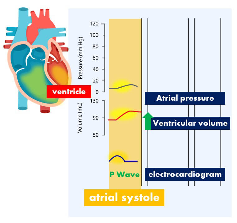
Right there, that bump in the volume of the ventricle. So this entire section is what happens during atrial systole. And we are ready for the next step.
Isovolumetric Contraction
Looking again at the ECG, we see that the next thing that happens is that we get the QRS complex. The QRS complex shows the depolarization of the ventricles. You can see, it’s much larger than the P wave and that’s because it’s a much larger structure – the signal is going to be larger. And just like what happened with the atria, with the ventricular depolarization, you expect to have ventricular contraction.
This is the beginning of the phase of systole, where the ventricles are contracting.
When they contract, what will that do to the pressure in the ventricles?
Well, during atrial contraction we did get a little increase in ventricular pressure of course – because blood was rushing into the ventricles, but now that the ventricles are contracting, you’re gonna see a much greater increase in ventricular pressure.
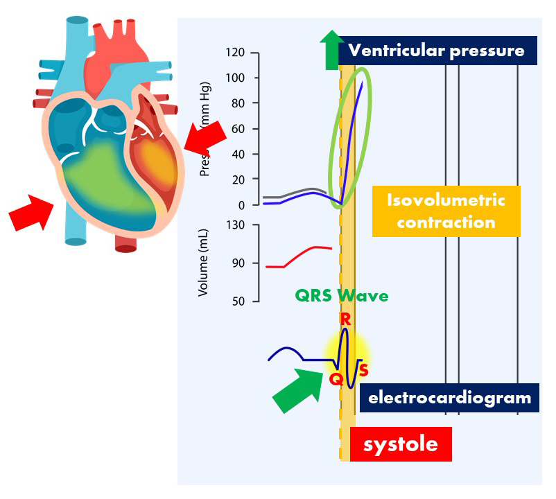
And it makes sense. You have a container, you squeeze that container, the pressure inside the container increases. In this case, the container is the ventricles, it’s made up of pretty strong muscle, so we get that huge increase in pressure.
Now, there’s a key thing that happens when the ventricles contract. As you see here, there’s a short phase called isovolumetric contraction. What exactly is that?
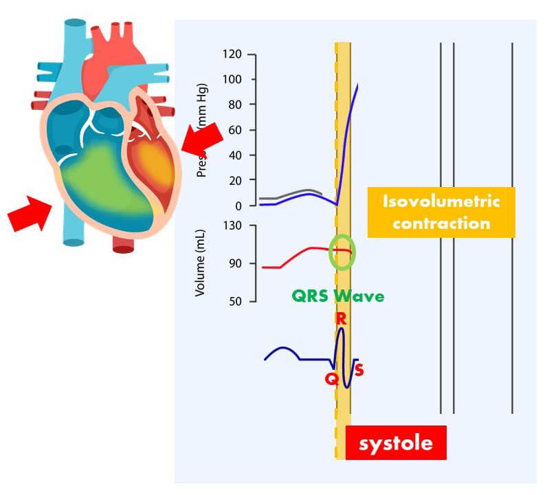
Well, the word isovolumetric means – the volume stays the same. The amount of blood in the ventricles remains the same. Look at the ventricular volume – it’s pretty much a straight line. And that’s because when the ventricles start contracting, that actually closes all the valves.
For example, if we’re looking at the left side of the heart, this atrioventricular valve gets shut and the semilunar valve is also closed.
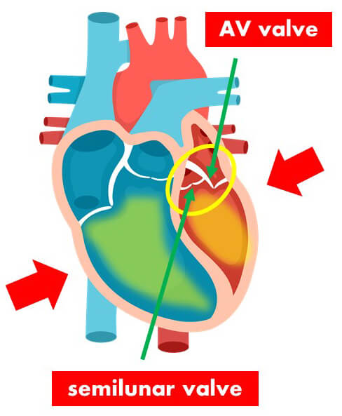
If they are both closed, we have a sealed container that is contracting, so we get this huge increase in pressure, but that isovolumetric stage only lasts a short period and that’s until the semilunar valve opens. That’s the valve between the left ventricle and the aorta. It’s going to be closed until a certain point.
Ejection Stage
Now, what point would that be? Well, let’s think about it.
On the other side of that semilunar valve is the aorta. And at this point right here, the aortic pressure is somewhere around 80 millimeters of mercury. So if you want to push blood in there, you have enough pressure in the ventricle to overcome that 80 millimeters of mercury.
And right at that point, the semilunar valve will finally open and the blood can be sent into the aorta so that it can go to the rest of the body.
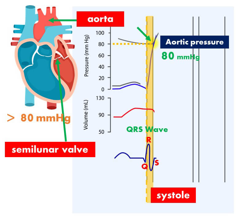
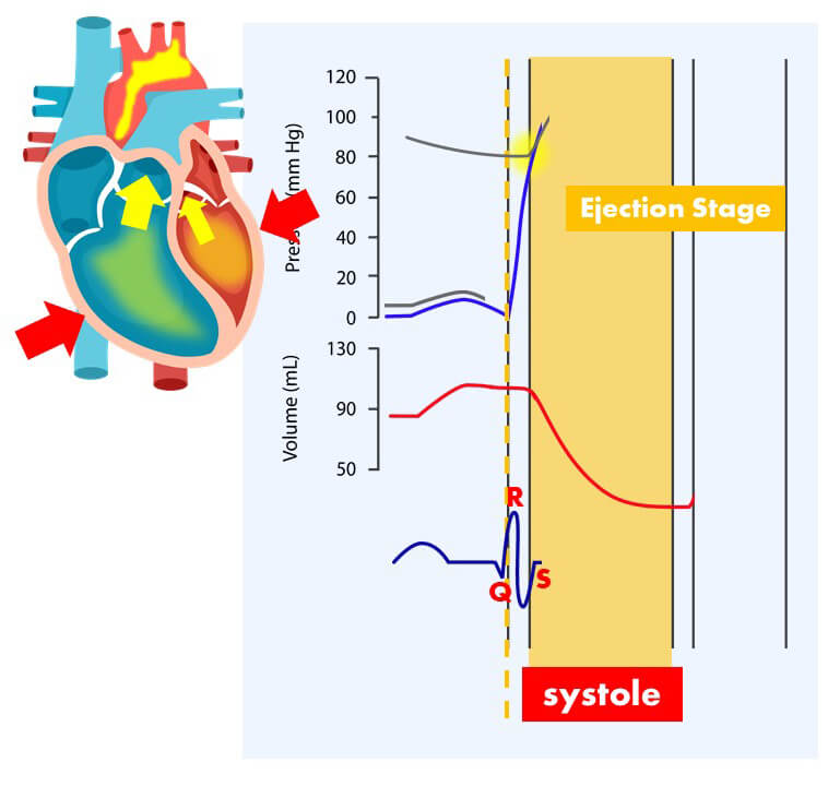
So, blood is leaving the ventricle. What will happen to the ventricular volume? It’s going to go down because we have blood getting out. And that’s exactly what you want. You want the blood to leave the ventricles and go out to the body.
This stage here is the Ejection Stage. That’s when blood is being ejected from the heart and specifically the ventricles.
Isovolumetric Relaxation
Let’s look back at the ECG, we then have the T wave. What does the T wave show?
Well, that’s Ventricular Repolarization – the opposite of depolarization. So the ventricles are going to relax now. What happens when the ventricles relax?
The pressure in the ventricles will come back down. At a certain point, the valves are going to close again and we get Isovolumetric Relaxation.
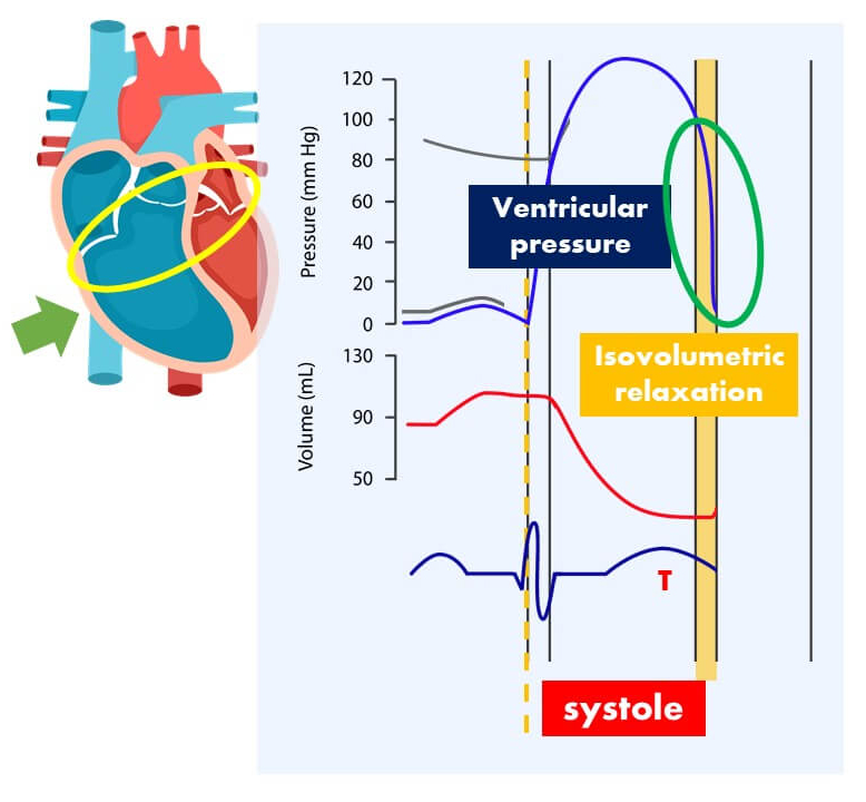
Valves are closed, ventricles are relaxing so the pressure in the ventricles drops significantly. And that’s exactly what you’d expect.
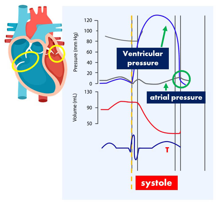
Now, once that ventricular pressure gets below the atrial pressure, what’s going to happen to the atrioventricular valve?
It’s going to open up again. And at that point, the valves are open and the blood that’s coming back from the body will just start passively filling the ventricles. And that continues up until the point where we get our next P wave to start the entire process again. That’s pretty much the cardiac cycle.
Phonocardiogram – Heart Sounds
Now, there’s one more thing that we didn’t cover and that is the phonocardiogram. That shows the sounds of the heartbeat. When you listen to the heartbeat, like with a stethoscope, you hear a sound that goes like this – lub dub. Lub dub. Lub dub. That’s what you’re seeing here.
What’s causing the Heart Sounds?
They are actually the sounds of the valves in the heart closing. Let’s look at when they happen. The first one happens right by the QRS complex.
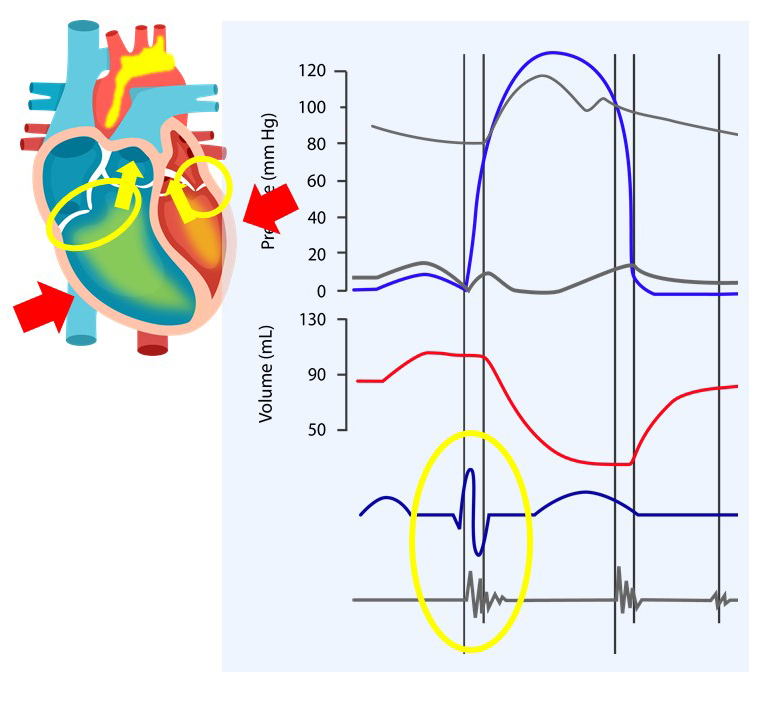
Remember, that shows ventricular depolarization, which causes the ventricles to contract. When the ventricles contract, that pushed the atrioventricular valve close. That’s why you get the first sound.
The second sound happens after the T wave – the ventricles relax, and the semilunar valves close. That causes the dub sound. That’s why we hear lub dub, lub dub, lub dub.
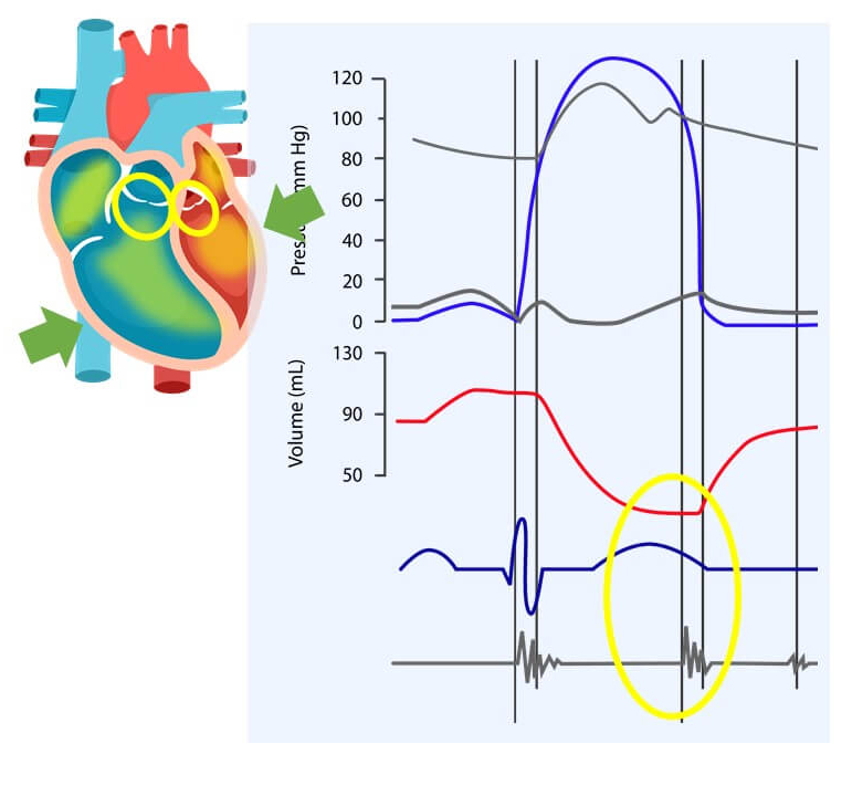
And now THAT is the entire cardiac cycle. Does it make sense?
If it doesn’t, watch it again, pause it where you need to, and get a good understanding of what this entire graph is trying to show.

this vid make cardiac cycle very clear to me and can you post vid talking about cardiac arrhythmia and i will be grateful
Unfortunately, I can’t. I’m focusing on making videos as I need them for my classes, so I can’t take anymore requests at the moment.
never mind,i appreciate that and all your vid are useful to me because i am in college of medicine
@ArwA1992 Oh wow, that’s pretty cool. Glad it’s useful!
This was so helpful, cheers!
Great video!
Question: Aortic valve opens when contracting vent. pressure exceeds the resting diastolic pressure in the aorta (80mmHg). Pressure waves cross there. At the end of vent. syst., relaxing vent. pressure drops below chock-full-O-blood aortic pressure and the valve shuts as aortic blood presses against the cusps. Why are the pressure waves different there? Why is aortic pressure 100mmHg and vent. pressure 60mmHg? Why doesn’t the aortic valve close at 80mmHg?
All questions are answered in the Interactive Biology forums from now on. Go to the website in the description and then visit the forum. This is to make it as efficient as possible as we have multiple people over there to help answer questions.
All the best
@kerrie337 Glad to know it helped. All the best!
awesome
you really made it fun not like our prof. in collage, keep up the nice work
@ToRnAdO0504563168 Thanks for the feedback. Glad you are enjoying it! Stay tuned for many more!
THank YOU!!
Thanx alot…..
Very helpfulll…..
Thanks a lot
Thank you!
thank you so much for helping 😀
@combymanje123 You’re welcome! PLease stay tuned because we have more Biology videos coming very soon!
@Shieek3080 You’re welcome. More biology videos coming soon!
@viju345 Glad you’re finding value in it. Visit our site for more fun Biology videos!
@miiigoreng You’re welcome! Stay tuned for more 🙂
@xty070 You’re very welcome! Stay tuned. New Biology videos will be uploaded soon!
You’re very welcome! Stay tuned. New Biology videos will be uploaded soon!
@ne0rne Thank you!
Remember walking into class late with all this stuff written on the board. i was so lost… not anymore. thanks a bunch.
@MaficSun You’re welcome! Stay tuned. We have more Biology videos to be uploaded very soon! 🙂
You’re welcome! Stay tuned. We have more Biology videos to be uploaded very soon! 🙂
A Little slow for my taste. Less intro, more info.
@Neopluto We’ll see what we can do. 🙂 Thanks for watching though! There are more videos you might want to watch from our Biology website. More will be uploaded so, please stay tuned!
We’ll see what we can do. 🙂 Thanks for watching though! There are more videos you might want to watch from our Biology website. More will be uploaded so, please stay tuned!
Great job sir.. Please keep continuing your great work. God bless you..
@WTF919ful Thank you and God bless you too 🙂
Thank you and God bless you too 🙂
Thank you Thank you Thank you :’)
@jadsal92 You’re welcome! Please stay tuned for more Biology videos to be uploaded very soon!
You’re welcome! Please stay tuned for more Biology videos to be uploaded very soon!
Thanks leslie!
شكراً
log duh log duh log duh xD
Thank you, thank you, thank you!!!!
U R AMAZING!!!!!!!!!!!!!
This is awesome Thank you VERY much !!!
You really make biology fun. Excellent video! keep going on. Greetings from Dominican Rep.
Thank you for this awesome video on the cardiac cycle!! So much better than trying to get all this from a textbook, and the zooming/video overlays are really useful!
Thank you for this awesome video on the cardiac cycle!! So much better than trying to get all this from a textbook, and the zooming/video overlays are really useful!
Thank you so much!! soo helpful
thanks alot.
man u r star !!!!
man u r star !!!!
omg! omg! omg!…ur just too awesome!!..if u were my teacher then i definitely would top every exams:)..thanks a lot
why the black mouse cursor eh?
This video gets to the point and is described clearly; did not over exaggerate or over complicate things either. I appreciate this video (and many other students) very much, thank you!
This video gets to the point and is described clearly; did not over exaggerate or over complicate things either. I appreciate this video (and many other students) very much, thank you!
Just saying,, the girls is HOT,, btw Thank You Very Much,,it helps,,, *whos the girl again?? Nvm
Just saying,, the girls is HOT,, btw Thank You Very Much,,it helps,,, *whos the girl again?? Nvm
great video… lubdub!! lubdub!!
great video… lubdub!! lubdub!!
great video… lubdub!! lubdub!!
Hi, Leslie Samuel! i really really like your Episodes. it’s very helpful for me. actually i’m a Tibetan monk who is learing homan physiology at Emory university. and my english is not good. so your videos with lecture article help me to good understand what’s going on in the classes. your speech is very clear and more easier to follow. thank you very much. i will be your student who really like teacher as you.
superb !helped me a lot.Thanks a lot.
superb !helped me a lot.Thanks a lot.
thankss
thankss
good stuff..
good stuff..
Finally I got it. Thanks a lot IB. I’m subscribing 2 ur site right now
Finally I got it. Thanks a lot IB. I’m subscribing 2 ur site right now
thank u very much sir….
thank u very much sir….
thank u very much sir….
thank u very much sir….
thank you for your sharing
thank you for your sharing
I wish it was you I was paying thousands of dollars to teach me this, instead of an overeducated old man who has tenure and can’t be fired from his university. I learned more here than I have in the passed 3 lectures. Thanks!
I wish it was you I was paying thousands of dollars to teach me this, instead of an overeducated old man who has tenure and can’t be fired from his university. I learned more here than I have in the passed 3 lectures. Thanks!
Thanks a bunch !
Thanks a bunch !
Thank you very much! Your explanation of cardiac cycle is much better than both Guyten&Hall and Ganong. Helps me very much in my preparations for the physiology exam in medical school in Norway.
Thank you very much! Your explanation of cardiac cycle is much better than both Guyten&Hall and Ganong. Helps me very much in my preparations for the physiology exam in medical school in Norway.
im watching all your muscle and heart videos one day before my finals and i must say iv learnt more in these past two hours than i have in the past 4months!!!!! THANK YOU
im watching all your muscle and heart videos one day before my finals and i must say iv learnt more in these past two hours than i have in the past 4months!!!!! THANK YOU
im watching all your muscle and heart videos one day before my finals and i must say iv learnt more in these past two hours than i have in the past 4months!!!!! THANK YOU
Someone may already have made this point, but I notice that in the diagram here the pressure in the ventricle drops below that in the aorta well before the aortic valve closes. I think a corrected image is on wikipedia now.
Someone may already have made this point, but I notice that in the diagram here the pressure in the ventricle drops below that in the aorta well before the aortic valve closes. I think a corrected image is on wikipedia now.
A-Level Biology students with BY2 tomorrow say thanks!
A-Level Biology students with BY2 tomorrow say thanks!
Merci Beaucoup!
Merci Beaucoup!
The best explanation ever!… Thumbs up for u man!
The best explanation ever!… Thumbs up for u man!
wow thank you hey, best explanation ever
wow thank you hey, best explanation ever
Thanks so much Leslie! I learnt so much on the cardiac cycle 😀
Who else was perving on that woman at 11:04 LOOOOOOOOOOL
Who else was perving on that woman at 11:04 LOOOOOOOOOOL
This was extremely helpful. I’ve watched it several times. THANK YOU!!!!!
This was extremely helpful. I’ve watched it several times. THANK YOU!!!!!
Thank you so much, you are a big help !
Thank you so much, you are a big help !
makes it crystal clear…you should be teaching at my college!
makes it crystal clear…you should be teaching at my college!
makes it crystal clear…you should be teaching at my college!
Hi Leslie congratulations and thank you, I just discovered your videos, I was thinking that another way that you can help you to afford this effort is become a partner with google and youtube, and allow ads in your videos, with any click to the ads that you got you will receive some money. I know you are doing this for share and help but also you have to be compensate for your time because is the only way to make this nice effort sustainable.
Thanks again and congratulations, i wish you success.
great explanation
great explanation
can you please post link for vector analysis ..i cant find it :/
Thank you for your time, you have a noble heart 🙂
totally awesome and your voice is so smooth and easy to listen too, no jumping around , concise all the way through. WOW!!! is right , so glad I saw this and your other videos. Give yourself a you tube nobel prize.
I seriously love your videos! Thank you for doing this! You are such a great help to students and anyone who just wants to learn everywhere!
This is amazing! Thank you so much. 🙂
Hey, your website and your video are awesome. I m a visual learner, and your videos help me perfectly with A&P class. Thanks a lot!
Genial Leslie!, saludos desde el sur
Great Leslie!, greetings from the south
They is WAAY simpler than how my teacher explain. Thank you.
Thank you soooo much!!!! This helped clear up some confusion before my exam!
You are Amazing!!!!!
Best video on cardiac cycle, hands down!
.k
Your such a great teacher,thanks a milloin 🙂
Your such a great teacher,thanks a million 🙂
I needed a quick refresher and this really helped. Best video I’ve seen on the subject.
Awesome explanation, especially right before my midterm for A&P!!
thank you for the video!! I understood so much more from this than a two hour lesson from my teacher ;w;
This video is great. Very detail!
I love your accent!!!!
Are you Jamaican?
I hear a hint of that in your accent.
Anyway great videos and greetings from Jamaica!
Thank you. great help! 🙂
No other video or college professor has been able to make this any more clear for me…and you did it in less than 14 minutes! I wish I would have found your videos earlier! This video makes the whole process so easy to understand. Your videos make an excellent reference. Thank you!
These videos are amazing! Thank you!!!
Fabulous video. You are a great teacher!!!
Thanks for your time may God bless u more.
I learned more in this 13 min then i did in 3 hours of lessons.
I think your speed is great. It allows me time to digest the information. Great job!!
thank you, you saved me. I wish my professor is as good as you 🙂
I’m a bit stuck with polarization can anyone provide a link to an explanation please
amazing teacher!!! thank you
Thank you so much!
I just discovered your channel and it is so helpful! 😀
Magic!
Congratulations and thank you very much for sharing all these videos. You make something complex very simple and clear !!! Some people said that it is a little bit slow, but for me it has the right speed to make it comprehensible. Thanks again !!!
This is amazing!
Thanks for the video 🙂 5* I’ve got an exam on the 9th of January 2013 and though a video could help! Will be visiting your website and may tell my friends about it! Thanks again
your channel is amazing!!
thank you and much respect
This is amazing, thank you so much, might just make it through my exams now!
what do the “c” and “v” waves on the atrial pressure wave indicate?
You’re videos are extremely helpful! Thank you so much!!
my friend told me about your videos and these are awesomes!! thanks a lot 🙂
c wave is due to rise in the atrial pressure produced by sudden closure and bulging of the tricuspid valve into the atria during isometric ventricular contraction , while v wave is due to rise in the atrial pressure as well but here due to accumulation of the venous return while the AV valves r closed , that’s all i know 😀
I just came.
My heart has gone through so many rhythms since watching this video. Makes you think a little!
man i swear this man Leslie Samuel deserves an award
Leslie Samuel is god
thank u indeed , an easy and simple way of teaching , but wishing a richer scientific subject .
Thanks, a Lot Samuel. Kui very Nice. I had neuer unterstand that diagram but now ist cristall clear. Thanks
I have a similar diagram in my book as was beyond confused in trying to make sense of it and understand how all the different parts correlate with each other. This video was absolutely amazing at describing each little part, and it put everything together like a story. Very thorough and helpful!!
Thank uuuuuuuuu:)
Im in nursing school and i couldnt understand this and I was soo stressed. But now i get it finally…I want to thank you!!!!!!!!! YOU HAVE NOOO IDEA HOW MUCH THIS HELPED ! KEEP IT UP :))))
You are absolutely amazing!! Can’t thank you enough
Thank you from the bottom of my heart! (corny pun intended) 🙂
I just discovered your channel. Great presentation with easy to absorb descriptions and instructional materials. I shall explore your other videos. Thank You!
I want to punch my professor -_- He gives me headaches!. You rock!!!!!.
Thank you!!!!! 😀
I just got a decent grade in my last anaotmy exam! Thanks soo much to this channel !!! I have another exam comming up and this vid is awsome!
Sir, you are AMAZING!!! Thank you for making this very easy and fun to understand!!
Come teach physiology at my school!!
Thank you for such informative, and easy to understand videos. You really do a wonderful job. Keep it up.
Excellent teaching of the Cardiac cycle, may God continue to bless you with your great generosity
Universities should replace all professors with these youtube personnel (people who actually enjoy teaching and speak proper English).
Comer pan negro, cereales integrales, frutas y lechuga. La leche de vaca es peligrosa. Tener alegria, habitos positivos y menos estres, enfrentar los problemas con esperanza y optimismo. No consumir grasa animal como la crema, helado de crema, etc. Recomiendo no fumar no tomar alcohol. La depresion hace mal, debemos buscar buen apoyo. Si se puede, realizar ejercicio aerobico. Llevar una vida espiritual y tener un caracter manso. Tomar mas agua conservando la moderacion en todo:.
Awesome, very clear and understandable language. It will helpful for me for my tomorrow physiology test. I learn much more in this 13 min than my hoursssssssss study.
Thank you, love it.
Thank you SO MUCH for these videos, I have a particularly inconsistent and confusing teacher for physiology who is unable to organize the material in a way that makes sense and is cohesive. I wish I could have paid you the tuition instead as these videos are how I am really learning. I wish all teachers could teach the way you do. you are amazing! Truly, from the bottom of my heart, thank you!
Great video, by the way it’s pronounced systole, like lamp pole. Not systoly
I would to thanks you for contributing time and efforts to make this video! A+++ Awesome! You are much better than my professor, who should learn more of how to teach! Not just speak!!
I would to thanks you for contributing time and efforts to make this video! A+++ Awesome! You are much better than my professor, who should learn more of how to teach! Not just speak!!
No, it’s not. He’s pronouncing it right.
Mulahemum YOUR AWESOME!!!!!!
Why does atrial pressure rise slightly during systole? Answer anyone?
because it contracts thus increasing the pressure within it,,and of course it will not contract as much as the ventricle contraction ( the blood will pass from the atrium to the ventricle below it,,so we don’t need a huge pressure to do that, bearing in mind that the flow of blood will pass passively at first depending on the pressure gradient only),,if you are asking about why does it increase in the ventricle systole,that is because the AV valves bulge into the atria…good luck 🙂
100% agree
Thank you 🙂
Also i love the way you say p wave! Your voice is amazing lol!
THANK YOU 😀
Thanks, this was very helpful.
This is even better with automatic captions on, haha
GREAT GREAT, THANKS <3
Am I the only person who sees a big blue penis on the right heart?
GREAT!
Thank you for this great video, I got an A in the lecture exam.
What a incredible explanation..2 thumb
i had never understand the cardiac cycle and how it works but now its diffrent i really wanna thank u
thnx alot
Thank You Internet!
Excellent!
I’ve found my new bestie!!!! Hello A. Thank you Sam. So clear, so concise, so simple.
Thank you?, very well executed!
I’m just wondering, when the ventricles are contracting and the aortic value is open, aka when the ventricular volume is dropping, are the atria filling up with blood?
I’m just wondering, when the ventricles are contracting and the aortic value is open, aka when the ventricular volume is dropping, are the atria filling up with blood?
Who ever gave this a thumbs down should go kill themselves
16 people,never went full retard.
I will love to have access to your program
Hi mr.samuel
these 3 questions have baffled me for a while,
Q1:when exactly dose the sodium-potassium pump’s job start?
1 right after the hyperpolarization 2 as soon as the cell reaches the resting state
if the assumption #2 is correct then in that case the hyperpolarization is sending K+ out while the pump is pumping K+ in?? is’nt it contradictory or maybe the assumption #1 is correct.
(plese check out my next comment because youtube doesnt let me to post it all in one comment)
Q2:IS refactory period the same as the activity of sodium-potassium pump? if the assumption 1 is correct then it can not be said that all that part of the graph which is under the line of resting state is all”refractory state”
Q3:why the Q and S in ORS complex are under hre isoelectric line?and which one causes the U wave, the depolarization of the purkinje fibers or repolarization of them
I would mean a lot if you take your time answering my question,thanks….
I love you … Thank you very much
I am now One Happy Med Student!!!
Thanks so much. I go to one of the best schools in UK but I understood better with this vid.
Why does the QRS wave go below the baseline? I am confused about what the axes of the graph are representing. What is on the y axis? Is going above the baseline becoming more positive? Or is it measuring volts or something of the like? Please help this poor confused student!
Hi, why is the volume in the ventricles increasing before you reach the Pwave? Is there blood entering the ventricles BEFORE the atria contract?
Yes you are correct. The ventricle AV valves are open and at ventricular low pressure (after relaxation) so the blood will pour in from the atria. Remember you have all that blood coming into the atria from the Vena Cava which makes atria have more pressure than vent. In young people ventricles actually get over 80% of the blood before the atria contract. When you get older however the ventricles get stiffer so that atrial pumping mechanism becomes more important to squeeze that blood in.
I can’t say anything more than: “Respect.” . I do respect this man for what he is doing and I think we all should.
Thanks for the video! Very helpful 😀
Life saver! :))
I love you!
Hey, from what I know, after the atria filling (isovolumetric relaxation), the pressure rises in atria causing the AV valves to open and blood starts filling the ventricles. THEN the atria depolarizes and contracts finish filling the ventricle (sort of like topping it off)
Thank you so much.
Haha, WIGGER
Haha, WIGGER
i wonder why some people will unlike this.
Thank you .
What word that was used is not considered english. Maybe you should teach people. Judging those who help, why?
I’ve learned more from you in just one video than I have from a human physiology class in a four year university. Thank you soo much! 😀
Thnx man !
Amazing Vedio.. it’s really helpful !!
Hey, Leslie. I’m a senior in college and am in Animal Physiology this
semester and it’s killing me! Thank you so much for taking the time to post
all these videos. They are so helpful!! I just wish I had discovered these
earlier and I’d probably have an A and not a B! 🙂
I love this!! Can I jump on this band wagon and help out???
Lub dub, lub dub! 😀 Thank you very much! This has been very helpful! 🙂
YOU ARE AMAZING ! i get it now 🙂
thank god for this
You explain things much more clearly than university lecturers in the UK! Thank you 🙂
I owe you my degree! Thank you 🙂
Amazing. Thank you!
I would like to add extra clarity to one commonly tested point: the heart sounds (seen on the phonocardiogram) are caused by blood hitting a closed valve. It is easy to think that the sounds are due to the valves themselves closing, but it is not so. Immediately after the valves close, blood smashes into the newly closed valves causing turbulent flow. It is this hitting of the valves and subsequent turbulence that results in the heart sounds. Mr. Samuel was not incorrect in what he said- I just wanted to emphasize that point. Thanks.