The early skeleton of a fetus is composed of cartilage and connective tissue. As the fetus grows these components are then replaced by bone, a process called ossification. This process can take many years to complete. There are two ways in which bones are formed:
- Intramembranous ossification
- Endochondral ossification
Intramembranous Ossification
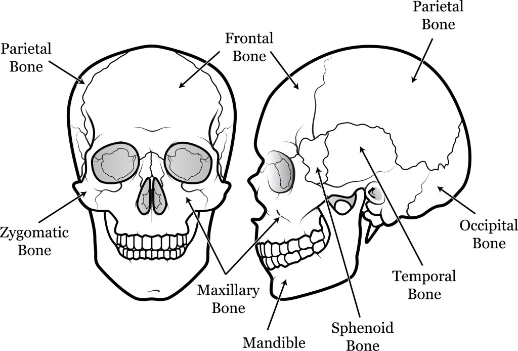
In intramembranous ossification models of bones are formed from membranes in the fetal skull and it results in the formation of cranial bones of the skull. These are:
In other words, flat bones are formed by intramembranous ossification. On the other hand most of the bones in the body are formed from endochondral ossification. It begins in the second month of fetal development and it is the process by which hyaline cartilage is being replaced by bone.
Endochondral Ossification
- Hyaline Cartilage is surrounded by perichondrium.
- This perichodrium is produced by chondroblasts.
- In the mid region of this hyaline cartilage which is covered by perichondrium, the cartilage calcifies.
- A primary ossification center is formed and a periosteal capillary grows into the calcified cartilage and supply its inside. The periosteal capillary along with bone forming cells (osteoblasts) then forms a periosteal bud.
- The periosteal bud invades the internal cavity and spongy bones are formed
- The shaft of the bone ossified from the primary ossification center, forming the diaphysis which continues to grow as the bone develops
- As the bone continues to grow secondary ossification centers will appear at various parts of the developing bone.
- From these secondary ossification center develops the epiphysis. A medulllary cavity also forms in the diaphysis.
- The epiphysis and diaphysis is separated from each other by the epiphysial plate.
- As the bone continues to grow, these plates are eventually replaced by bone and bone growth ceases and the diaphysis then fused with the epiphysis. This occurs between ages 18 and 25 years old.
Parts of the long bone

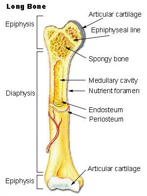
- Epiphysis –this is the extended part of the end of the bone. One could say the wide end of the long bone and it is very important in bone growth. It is composed mainly of spongy bone.
- Diaphysis – this is the shaft of the long bone, it is covered by periosteum and contains compact bone
- Epiphyseal plate (of cartilage) – covers the epiphyses of bone.
- Metaphysis-this is the flared part of the diaphysis and its closest to the epiphysis
- Medullary Cavity– this is a hallow space between the shaft of lone bones and it is filled with bone marrow
- Periosteum – Outermost layer that supplies blood and nerves to the bone
- Compact bone -hard bone beneath periosteum mainly found in shaft
- Spongy bone – this bone is light and is found near joints
- Bone marrow – there are two types: Red and yellow bone marrow.
Red bone marrow found in children and adults, it is required to make red blood cell. Yellow bone marrow is yellow and is filled with fatty tissues.


And as an extra tit-bit of information, red marrow turns to yellow marrow as you get older however, in times of need such as severe blood loss, the yellow marrow can revert back to red!