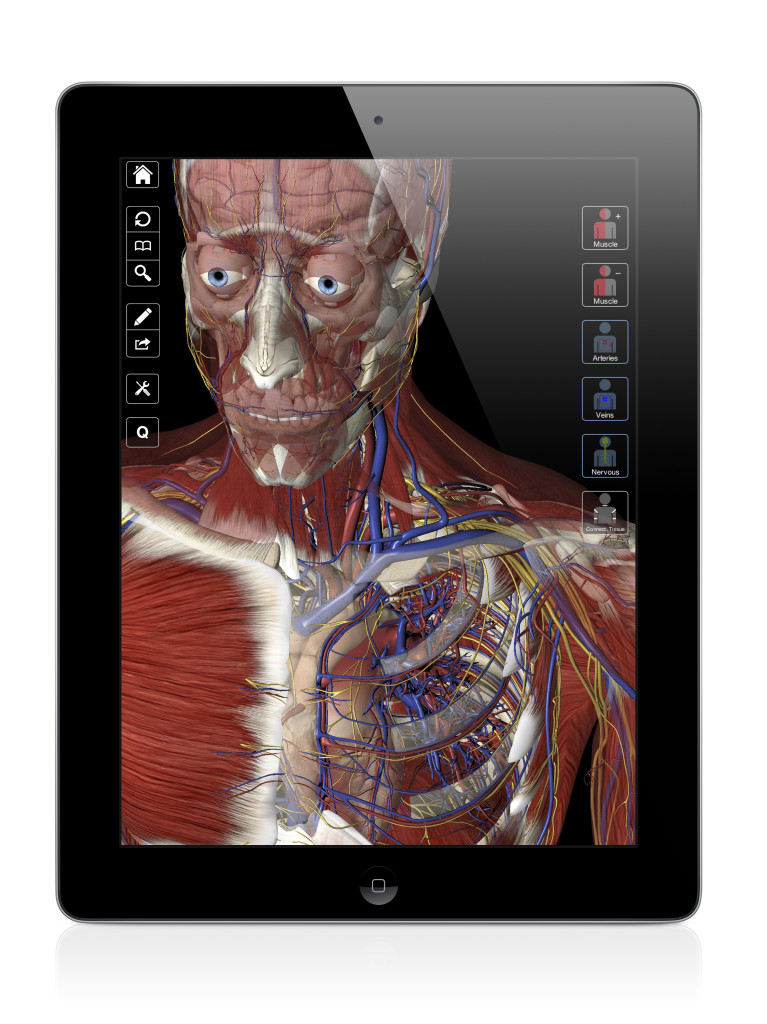
In this video, Leslie talks about the different boundaries of the Cubital Fossa. What are they? Watch to learn more.
Enjoy!
Transcript of Today’s Episode

Hello and welcome to another episode of Interactive Biology TV where we’re making Biology fun. My name is Leslie Samuel and this video is brought to you by our partners over at 3D4Medical.com, the creators of this app which is called Essential Anatomy. They have created this one for the iPad and you can get it in the App store and they have a number of other apps. This once again is called Essential Anatomy and you can find it in the App store.
In this video, I’m going to be talking about The Boundaries of the Cubital Fossa. Let’s get right into it.
The cubital fossa is this little triangular depression that you see right here in the region between the two forearm bones. So, you have the forearm bones — the radius and the ulna.
The radius, of course, is going to be lateral and the ulna is going to be medial. So, just imagine, the two bones that are under here, the radius and the ulna, and right in between those two, right as you go from the arm to the forearm region, you’re going to get a triangular depression. Let me actually zoom in a little more. There we go. That’s good.
You can actually feel this. If you just took your fingers right now and put them between your arm and your forearm, you’ll feel this little depression that I am going to outline right now.
Right here, you’ll see this little triangular depression. It’s shaped like a triangle with the base that’s superior. So, this is your superior base. And then, of course, you have the other two sides of the triangle.
So, first, I want to talk about what those boundaries are. First, the base is going to be this line that you see right here. Now, there isn’t any particular structure that outlines that base. So, that’s kind of like an imaginary line between the two epicondyles.
All right, so we have the lateral epicondyle here which is this part on the lateral side. When you look at the structure of the humerus, you’ll see we have the lateral epicondyle and of course, we have our medial epicondyle which is the larger of the two epicondyles. So, I should have drawn that as a bigger bulge.
So, we have the medial epicondyle and the lateral epicondyle. And, there is an imaginary line that we’re imagining right between those two structures. That’s the base of the triangle.
Then of course, the lateral border of that triangle is going to be this muscle right here. That’s your brachioradialis muscle. Then, the medial border, that’s going to be this muscle right here, the pronator teres muscle and that is going to give you your entire triangle — from the base being that imaginary line between the two epicondyles then, we have our lateral boundary which is made up of the brachioradialis muscle and the medial boundary is outlined by that pronator teres muscle.
Then, of course, we have to have a roof and a floor. Let’s take the floor first because we can see that right here. In order to see the floor, I’m actually going to hide your biceps muscles and then, here, you can see this brachialis muscle which is going to be forming the floor of that triangle.
So, that’s the floor of the little depression that you have there and then, of course, we can’t have a floor without having a roof. That’s just not as cool. So, let’s add our roof and the roof will be formed by a number of structures.
This first structure that you see here is the bicipital aponeurosis. You have the biceps that are inserting on that… Well, they’re using that bicipital aponeurosis which is a flattish tendon. It serves as part of the insertion of the biceps muscle, the biceps brachii. That’s the first part of the roof.
The second part has to do with a vein that’s going through that cubital fossa and that is your median cubital vein, that vein that I have highlighted right there.
Then, the third aspect would be two nerves that we have coming through. I’m just going to draw them in but, you have a medial and a lateral cutaneous nerves of the forearm that makes up also part of the roof of that cubital fossa.
So, in review, let’s look at all of that again.
When it comes to the boundaries, we have our base which is formed by that imaginary line between the lateral and medial epicondyle. You have your lateral boundary which is formed by your brachioradialis muscle. You have your medial boundary and that is formed by the pronator teres muscle. Then, you have the floor, I’m not going to show that right now, but the floor is made up of the brachialis muscle and the roof, we have our three structures — the bicipital aponeurosis, the median cubital vein and then, the two nerves, the lateral and the medial cutaneous nerves of the forearm.
So, that’s pretty much it for this video. If you want more videos like this and other resources to help make Biology fun, you know what to do. Head on over to the website, interactive-biology.com.
This is Leslie Samuel. That’s it for this video and I’ll see you in the next one.
[table “” not found /]
Thank you for such cool videos.They are great for revision 😀
Hi! just wanted to say thank you for these videos
Anatomy is my weak point and I always get so overwhelmed with all the information
thanks to your videos I feel like I understand better and it is more enjoyable 🙂
I just wish that I would have found your videos earlier!!